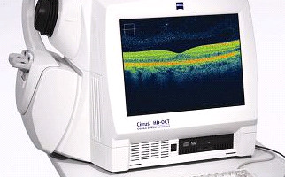
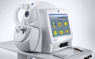
OCT Cirrus HD coherence tomography
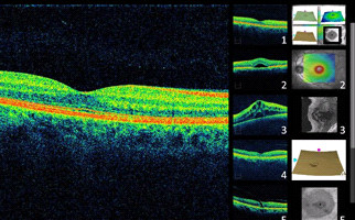
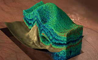
Optical coherence tomography OCT Cirrus is a modern tomographic imaging method of the retinal structure, allowing examined tissue’s optical biopsy for diabetic retinopathy, aged-related macular degeneration, macular hole, cystoid macular edema, central serous choroidal retinopathy and glaucoma cases.
Cirrus HD-OCT system is based on spectral (spectroscopic) optical resolution technology (Spectral Domain OCT). Spectral domain OCT technology produces retinal structure images and optic papilla morphology using special analysis. The only certainty is that patients who undergo spectral optical coherence tomography with Cirrus HD OCT, they receive fast, reliable, analytical, qualitative and documented examination results.
The smallest pupil diameter required for measurements, among competition (≥ 2mm), GPA software for glaucoma patients complete monitoring, ergonomic design for minimum space occupation, Line Scanning Ophthalmoscope (LSO) for precise OCT and fundus image identification and also repeatable results, extremely easy operation, diagnostic and statistical software analysis, retinal layers separation algorithm, automated determination of macular fovea and optic papilla and high resolution pachymetry maps are some of the system’s key advantages.


