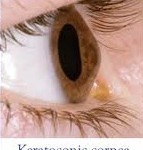


Keratoconus
What exactly is keratoconus and which are its causes?
Keratoconus is a corneal condition. More specifically, cornea’s anatomical shape is deformed, developing a conical swelling (not an inflammatory keratektasia), resulting in a dangerous thickness thinning. The exact etiology of the condition has not been clarified, but there are indications that heredity is one of the main causes. Keratoconus, in most cases, makes its appearance in puberty and develops over time. Usually, it affects both eyes (bilaterally). In advanced stage keratoconus can be detected even with the naked eye. Diagnosis of keratoconus should promptly occur, otherwise there are possibilities for a keratoconic patient to undergo even a corneal transplantation surgical procedure (keratoplasty).


Symptoms:
The main symptoms of keratoconus are the following:
- Decreased visual acuity
- Increased photosensitivity / photophobia
- Vision blurring and distortion
Keratoconus treatments:
In early stages of keratoconus eye spectacles and contact lenses are used to restore refractive problems (e.g. myopia or astigmatism). But soon such solutions are not too effective to cease keratoconus progress. Many patients use semi-rigid or rigid, semi-permeable keratoconic lenses.
In recent years, intrastromal corneal rings are used (ICRS – Intrastromal Corneal Ring Segments), in order to improve and normalize the topographic profile. The results are encouraging, but an important criterion is the suitable selections of cases.
Corneal transplantation (keratoplasty) surgical procedure (penetrating or lamellar) is recommended in advanced stages of keratoconus.


Collagen Cross Linking (C3R/CXL) technique:
A new and extremely promising therapeutic approach for facing and eliminating keratoconus is collagen cross linking technique, using riboflavin (B12) and ultraviolet (UVA) radiation (Corneal Collagen Cross Linking C3R/CXL). Via this method, the creation of new bonds between corneal collagen fibers is performed, resulting in corneal stiffness stabilization. So continuous corneal thinning is prevented and keratoplasty option is avoided.
During treatment, after local anesthetic use and part of epithelium (the outer membrane of the cornea) removal, riboflavin drops are applied to cornea, usually for half an hour and finally the keratoconic eye is exposed to UV-A (ultraviolet) radiation (while continuing riboflavin B12 instillation) for extra 30 minutes. At the end of procedure a protective contact lens is placed on the eye preventively.


It is worth mentioning that corneal collagen cross linking technique can be appropriately combined with excimer laser, more specifically with topo-guided (T-CAT) PRK (Photo Refractive Keratectomy) treatment method for simultaneous refractive error (diagnosed with keratoconus progress) restoration and vision recovery.